IDF logo
Helix

Complement deficiencies

Individuals with a complement deficiency can have clinical problems that are a result of the role that the specific complement protein plays in the normal function of the human body.

The complement system consists of more than 40 proteins, present in fluids (blood) and tissues, and other proteins anchored on the surfaces of cells. Most of the blood complement proteins are made in the liver, though a few of them are made by cells outside the liver, including neutrophils and fat cells (adipocytes). The primary functions of the complement system are to protect the body from infection, to remove particulate substances (like damaged or dying cells, microbes, or immune complexes), and to initiate and modulate (change) immune responses. As part of the innate immune system, the complement acts immediately to start the process of removal and resolution of the problem, such as an infection. The complement works with cells of the immune system and proteins of the coagulation pathway to control an infection. There can be deficiencies in any one of the many individual complement proteins. Individuals with a complement deficiency can have clinical problems that are a result of the role that the specific complement protein plays in the normal function of the human body.

Activation of the complement system and its pathways

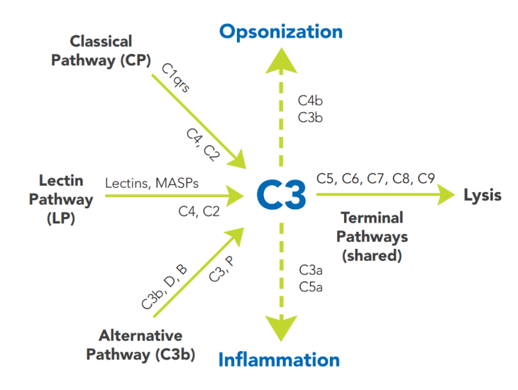
Complement activation is designed to eliminate invading microbes while producing minimal collateral damage. Complement proteins in the circulation are generally not activated until triggered by an encounter with a bacterial cell, a virus, an immune complex (antibody attached to a foreign material), or damaged tissue.

Complement activation is a cascading event with tremendous amplification potential. It must follow a specific order if the result is to be achieved. The circulating proteins have been grouped into three major activation pathways, based on the types of substances and proteins that initiate the activation. If you visualize a trident, the three tines (teeth) represent the different initiation routes of the complement system, while the handle represents the mechanism by which this cascade ultimately destroys the threat, no matter which activation pathway started the response. The diagram in Figure 22:1 depicts the activation pathways.

Figure 22: 1 Complement system and its pathway
 

Figure 22:1 Complement System and Its Pathway

The alternative pathway (AP) is the most evolutionarily primitive of the complement pathways. It is initiated by fragments of the complement component C3. These are always being generated at a low level in our body but get rapidly amplified when they encounter a pathogen or abnormal cells. Other elements of the AP are Factor B, Factor D and properdin. A unique feature of the AP is the presence of the only positive regulator in the complement system, properdin. Properdin makes it possible for the amplification loop of the AP to efficiently put lots of C3b onto the surface of the activating cells, bacteria, protein complexes, or particles in the immediate vicinity of the activation site. Because the ability of C3b to bind to these surfaces decays rapidly, the activation is limited to just the region around the C3 cleavage (activation) site. This time-limitation is another control mechanism for the complement pathway. 

The classical pathway (CP) is activated primarily by immunoglobulins (antibodies, including autoantibodies) that are bound to antigens, either in the fluid phase as soluble immune complexes or on the surfaces of cells. Components of the CP are C1q, C1r, C1s, C2 and C4. The CP was the first to be discovered, but is the most recent in evolutionary terms. 

The lectin pathway (LP) is similar to the CP except for the first two steps. Mannose-binding lectin (MBL), the ficolins, and collectins initiate the LP. These are often present on surfaces of pathogens or altered host cells (such as dead cells). C2 and C4 also participate in the LP. 

The terminal pathway (TP) is the final set of steps in the complement activation process that forms a membrane lesion or hole (membrane attack complex, MAC), thus killing susceptible pathogens (bacteria) or other cells that activate complement on their surfaces. The TP is dependent upon at least one of the other pathways, such as AP, CP, or LP for activation. The components of the TP are C5, C6, C7, C8 and C9, and they form the MAC (C5bC6C7C8C9, or C5b-9).

Regulatory proteins to prevent unregulated activity (and tissue damage) are present as control mechanisms for each pathway. These can be present in the fluid phase, such as blood, or on the cell surface. C1-esterase inhibitor (C1-INH) is a serine protease inhibitor (SERPIN) in the blood and acts by forming a complex with active enzymes to trap and inactivate them. It is important in controlling the C1r and C1s activation in the classical pathway and the MBL-associated serine proteases in the lectin pathway along with enzymes in the coagulation system. Factor H and Factor I are also fluid phase proteins that help to regulate the amplification of the complement system. Their deficiency can result in a condition known as atypical hemolytic uremic syndrome (aHUS), which is associated with red blood cell damage and kidney dysfunction. CD46 and CD55 are proteins present on the cell surface, that also limit complement activation on the cell from which they are expressed. CD59 deficiency is associated with lysis of red blood cells and kidney damage, as seen in paroxysmal nocturnal hemoglobinuria (PNH).

The dynamic interplay among the different complement pathways and their control processes involves other plasma protein systems such as enzymes of the coagulation system, enzymes from inflammatory cells, and substances such as histamine and elastase released from cells in the local environment. All of these participants affect the outcome of an activation event. Most of the time, the outcome is favorable to the host. The diseases that accompany uncontrolled activation or a defect in the function of complement are often the result of an inherited deficiency or subtle impairment of one or more of its regulators. 

Find complement deficiency clinical trials

See if you qualify to participate in clinical trials evaluating new treatments and/or diagnostics for complement deficiencies.

Complement deficiencies and their diagnosis

Over 50% of people who have a deficiency in complement proteins develop infections. An even higher percentage of individuals with a deficiency in the CP develop autoimmunity, primarily systemic lupus erythematosus (SLE). A deficiency of C3, the most abundant complement protein, results in severe, recurrent infections (especially those of the respiratory tract) that begin soon after birth. As the age increases, infections are primarily due to a bacterial species, Neisseria, which can cause meningitis. Hence, the typical clinical symptoms of complement deficiencies include recurrent mild or serious bacterial infections and autoimmune disease. In C1 inhibitor deficiency, episodes of angioedema (a swelling under the skin or in the intestines) occur. The list of potential complement-related health problems includes renal disease, vasculitis (blood vessel inflammation), and age-related macular degeneration. A history of family members having the same presentation should increase the suspicion of an inherited complement deficiency.

The initial tests done to evaluate an individual’s complement system can identify an inherited defect and indicate what further testing must be done to make the diagnosis. The aim of the evaluation process is to define the complement component deficiency, while ruling out acquired causes of low complement values. Several screening blood tests are available that make it easier to find the answers. These include CH50, which is a useful tool to screen for classical pathway deficiencies, and AH50, which is used to screen for alternative pathway deficiencies. Individual complement levels can also be tested. It is important to know as much as possible about the reason(s) for low or absent complement so that decisions regarding appropriate treatment can be made, including when to use antibiotics and immunizations as well as for decisions about genetic counseling for inherited deficiencies.

Therapies specific for complement deficiencies are still in the developmental stage for most components, but in some cases, such as C1- INH deficiency, there are currently a number of medications available. For poorly controlled complement activation, especially that occurring due to deficiencies in regulatory proteins as in aHUS or PNH, there are certain drugs available to treat acute episodes or to prevent recurrence. Additionally, a monoclonal antibody to C5 (eculizumab) is used to block complement-induced damage. Additional therapeutic agents may become available in the next decade.

Deficiencies in the alternative pathway: C3, factors D, B, and properdin

Inherited C3 deficiency results in repeated, severe infections starting from birth, especially those of the respiratory tract. Organisms such as Streptococcus pneumoniae and Hemophilus influenzae are the most frequent causes of infection. These infections have decreased in frequency and severity due to use of antibiotics and vaccines.

Properdin is the only complement protein that is X-linked, so its deficiency only affects males. The protein is synthesized by immune cells such as monocytes, granulocytes, and T cells. Properdin deficiency increases the susceptibility to bacterial infections of the Neisseria family of organisms. The most prominent in the group is Neisseria meningitidis, which can cause a serious form of meningitis. Typical family histories include male relatives who have had or died from Neisserial infections.

Factor D deficiency is very rare and has only been described in two families. Both of these families had multiple members with a history of serious infections.

Deficiencies in the classical pathway: C1, C4, C2, C1-INH

Rapid clearance of immune complexes, dying cells, and debris from damaged tissues is a job that is efficiently performed by a normal classical pathway. The C1 protein is made up of C1q, C1r, and C1s. Complete deficiency of C1, C2, or C4 is closely linked to the development of systemic lupus erythematosus (SLE). This is thought to be due in part to the inability of complement to clear immune complexes and debris from dying cells, especially DNA and RNA.

C2 deficiency is the most common complement deficiency in populations of European descent, with frequency estimates between 1 in 10,000 to 1 in 20,000 individuals are homozygous C2-deficient. In primary immunodeficiencies (PI), C2 deficiency is found in young children who have recurrent infections, mainly upper respiratory tract or ear infections due to Streptococcus pneumoniae and Hemophilus influenzae. Many adults with C2- deficiency also have SLE.

Hereditary angioedema (HAE) is a disease caused by deficiency of the CP control protein, C1- INH. Symptoms generally begin around puberty but can occur earlier. These individuals have recurrent moderate to severe swelling in the extremities, face, lips, larynx (vocal cords), and gastrointestinal tract (intestines) that is frequently painful and debilitating. Intestinal swelling can cause acute severe abdominal pain, and the acute intestinal swelling can result in obstruction, which may need acute surgical intervention. Swelling the vocal cords can be concerning because of the possibility of airway obstruction leading to suffocation. These episodes typically last 3-5 days if not treated with effective medication.

Acute treatments for HAE include C1 inhibitor, a replacement therapy [both plasma-derived (concentrate) and recombinant products are available]; ecallantide, a kallikrein inhibitor (available in the U.S. only) and icatibant, a bradykinin-2 receptor antagonist. Note that steroids and antihistamines are not effective to treat HAE or prevent attacks. Prophylactic treatments include androgens, C1 inhibitor concentrates, and lanadelumab.

Deficiencies in the lectin pathway: MBL, M-ficolin, L-ficolin, H-ficolin, CL-11, MASPs

Deficiencies in the lectin pathway are fairly common, affecting approximately 5-30% of individuals. There is some controversy over the importance of the lectins to overall immunity. Most experts agree that the lectin pathway is important to fight bacterial infections during the early months of a baby’s life when maternal antibodies decrease, and the child’s own antibody production is not fully functional. In certain cases, the deficiency may be severe. [For example, a homozygous mutation in the ficolin-3 gene leads to severe, recurrent pulmonary infections in children that can result in severe lung disease, including bronchiectasis (damage to the lungs), later in life. Other studies have shown increased susceptibility to HSV-2 (herpes simplex virus-2), influenza A, Pseudomonas aeruginosa, and Staphylococcus aureus. Homozygous mutations in either the collectin-11 gene or MASP-1 gene can result in developmental abnormalities (including facial irregularities, cleft lip and palate, and cognitive defects).

Mannose binding lectin deficiency

Mannose binding lectin deficiency (MBL) is a part of the lectin pathway of the complement system, one of several different components of our immune defense. The lectin pathway may be the first to react before a traditional immune response occurs. It was thought that deficiency of MBL might explain some cases of increased susceptibility to bacterial infection. However, when a test was developed to measure MBL in the blood, it was determined that low or absent MBL is very common, affecting approximately 5-30% of all individuals. Therefore, its absence alone cannot be a cause of serious immunodeficiency, or a large portion of the world’s population would suffer from frequent major recurrent and potentially fatal infections. The MBL test is occasionally still ordered during an evaluation for immunodeficiency, and when the results show MBL to be low or absent, it is wrongly interpreted as indicating the presence of a PI. By contrast, expert immunologists experienced in caring for people with PI believe that low or absent components of this lectin system, including low or absent MBL, do not cause immunodeficiency by themselves. There is no recommended treatment for low or absent MBL, and immunoglobulin replacement therapy is clearly not indicated for that purpose. It is important to stress that the finding of low or absent MBL does not indicate that the cause for an individual’s infections has been found and that the diagnostic process must continue until the correct diagnosis is determined.

Deficiencies in the regulatory proteins: Factor H, factor I, CD46, CD55, and CD59

Complete deficiency of factor H leads to uncontrolled activation of the alternative pathway and C3 consumption. Recent data has been published that demonstrates how critical the role of this complement control protein is in maintaining health in a number of tissues. In addition to bacterial infections, deficiency or dysfunction of factor H is associated with various forms of kidney disease, including atypical hemolytic uremic syndrome (aHUS), as well as age-related macular degeneration (AMD). Heterozygous variants in factor I and CD46 can also result in aHUS. These diseases are examples of control processes gone awry on the surfaces of the organs affected. An acquired deficiency of CD59 is seen in paroxysmal nocturnal hemoglobinuria, which is associated with hemolytic anemia and renal failure. More recently, genetic sequencing has identified mutations in CD55 that are associated with CHAPLE syndrome (complement hyperactivation, angiopathic thrombosis, and protein-losing enteropathy), which often presents as abdominal pain or diarrhea, recurrent infections, and thromboembolic disease (blood clots). 

Treatment of complement deficiencies

Any complement deficiency should be treated as a form of PI, and the individual should be immunized against the bacteria that are most likely to infect them (see above). For example, boys with properdin deficiency should be immunized against Neisseria meningitidis, and meningococcal vaccines in addition to the usual childhood vaccinations. People with deficiencies of the other alternative pathway components and the terminal pathway proteins are also susceptible to Neisseria meningitidis and should be immunized. Antibody responses should be checked after vaccination, since the inability to activate complement may impair response to the vaccine.

Currently, there is no single treatment for complement deficiencies. Appropriate prevention and treatment of infections (usually with antibiotics) is key. Fresh frozen plasma infusions have been tried in some cases but carry a risk that the individual may make antibodies to the missing complement component, so prolonged use is not advised. In the case of HAE, effective medications to treat or prevent angioedema episodes are available. In aHUS or PNH, eculizumab, a complement-inhibitory antibody, may help. Prophylactic antibiotics can be used if the individual experiences repeated infections. Most of these individuals who are predisposed to infections eventually make antibodies against the offending bacteria and do not get sick as often.

Summary

The inherited deficiency of various complement proteins results in diseases due to the inability to perform primary immune functions, such as protection from infection or clearance of cellular debris. Additionally, in most situations where autoantibodies are present (such as SLE), these antibodies can engage complement, consume it, and result in tissue damage. Thus, there are certain acquired deficiencies of complement that are not inherited but can lead to disease. Both children and adults can be affected by complement deficiencies, so it is important to recognize the typical presenting symptoms of specific deficiencies as outlined above. Knowledge of the role of each complement component in the body and how it is regulated can help in understanding the effect of the specific deficiency and its treatment.

This page contains general medical and/or legal information that cannot be applied safely to any individual case. Medical and/or legal knowledge and practice can change rapidly. Therefore, this page should not be used as a substitute for professional medical and/or legal advice.

Adapted from the IDF Patient & Family Handbook for Primary Immunodeficiency Diseases, Sixth Edition 
Copyright ©2019 by Immune Deficiency Foundation, USA