
-
Understanding primary immunodeficiency (PI)
Understanding PI
The more you understand about primary immunodeficiency (PI), the better you can manage it. Learn about PI diagnoses and treatment options.
-
Living with PI

Living with PI
Living with primary immunodeficiency (PI) can be challenging, but you’re not alone—many people with PI lead full and active lives. With the right support and resources, you can, too.
-
Get involved

Get involved
Be a hero for those with PI. Change lives by promoting primary immunodeficiency (PI) awareness and taking action in your community through advocacy, donating, volunteering, or fundraising.
-
Advancing research and clinical care

Advancing research and clinical care
Whether you’re a clinician, researcher, or an individual with primary immunodeficiency (PI), IDF has resources to help you advance the field. Get details on surveys, grants, and clinical trials.

Key points:
- Primary immunodeficiencies (PIs), also known as inborn errors of immunity (IEIs), are rare, chronic health conditions where part of the immune system is missing or does not work properly.
- Infections are the most common symptom of an immune system that isn’t working properly.
- Autoimmune conditions, chronic inflammation, recurrent fevers, and cancer can also be signs of immune system issues.
- Some PIs, known as primary immune regulatory disorders (PIRDs), also involve problems controlling the immune system.
- Secondary immunodeficiency (SI) happens when a condition or factor outside of the immune system affects how well the immune system works. SI is much more common than PI.
Who does PI affect?
A recent study of medical records estimates that about 1 in every 1,600-1,700 people in the U.S. have some form of PI with documented symptoms [2]. PI can affect anyone, regardless of age, gender, or ethnicity. Some PIs like WAS cause symptoms in babies or young children, but others, like CVID, may not show up until mid- or even older adulthood.

What is secondary immunodeficiency?
Unlike PI, secondary immunodeficiencies are caused by conditions or factors outside of the immune system that affect how well the immune system works. Secondary immunodeficiencies are much more common than PI and healthcare providers have to rule out secondary causes before making a PI diagnosis.
The most common causes of secondary immunodeficiencies are aging, poor nutrition, taking chemotherapy or other immune-suppressing medications, human immunodeficiency virus (HIV), and protein loss in the intestines or kidneys. It’s important to recognize these conditions because, if the underlying cause can be treated, immune system function usually improves. Managing a secondary immunodeficiency is often similar to managing PI.
Understand PI more clearly

How your immune system works
To better understand PI, it's helpful to know more about how your immune system works.
Read about the immune system

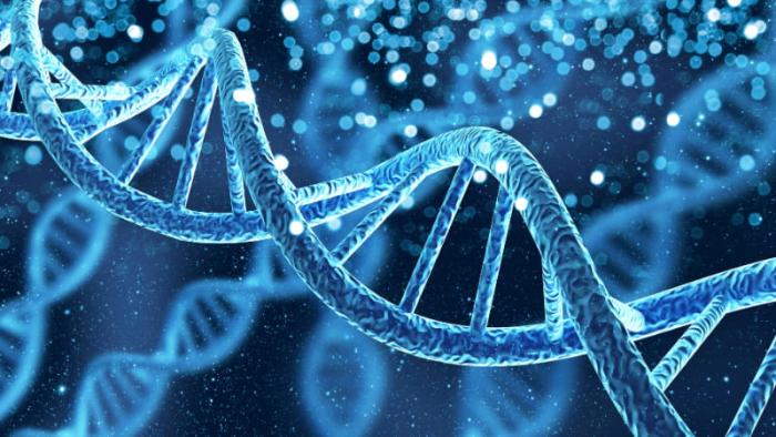
Genetics and PI
PIs are mostly caused by changes, or variants, in genes that are important for how the immune system works.
Learn about genetics
Types of PI
There are more than 550 primary immunodeficiencies with distinct definitions, causes, and symptoms.
Explore PI types
This page contains general medical and/or legal information that cannot be applied safely to any individual case. Medical and/or legal knowledge and practice can change rapidly. Therefore, this page should not be used as a substitute for professional medical and/or legal advice. Additionally, links to other resources and websites are shared for informational purposes only and should not be considered an endorsement by the Immune Deficiency Foundation.
Adapted from the IDF Patient & Family Handbook for Primary Immunodeficiency Diseases, Sixth Edition.
Copyright ©2019 by Immune Deficiency Foundation, USA.
Sign up for updates
Receive news and helpful resources to your cell phone or inbox. You can change or cancel your subscription at any time.