
-
Understanding primary immunodeficiency (PI)
Understanding PI
The more you understand about primary immunodeficiency (PI), the better you can manage it. Learn about PI diagnoses and treatment options.
-
Living with PI

Living with PI
Living with primary immunodeficiency (PI) can be challenging, but you’re not alone—many people with PI lead full and active lives. With the right support and resources, you can, too.
-
Get involved

Get involved
Be a hero for those with PI. Change lives by promoting primary immunodeficiency (PI) awareness and taking action in your community through advocacy, donating, volunteering, or fundraising.
-
Advancing research and clinical care

Advancing research and clinical care
Whether you’re a clinician, researcher, or an individual with primary immunodeficiency (PI), IDF has resources to help you advance the field. Get details on surveys, grants, and clinical trials.

Key points:
- The immune system is a network of many different organs, cells, and proteins throughout the body that work together to protect against threats like germs.
- The innate immune system includes all the immune system parts that don't need training, such as cytokines, natural killer (NK) cells, neutrophils, and monocytes/macrophages. Innate immune responses are not targeted to specific germs and include fever, inflammation, and swelling.
- The adaptive immune system includes the parts of the immune system that 'learn' to target specific germs, such as B cells, antibodies, and T cells.
- Both the innate and adaptive immune systems are present from birth, but the adaptive immune system develops rapidly during childhood as it gains experience with different germs.
- Different pieces of the innate and adaptive immune systems protect against specific kinds of germs like bacteria, fungi, or viruses.
Major organs and tissues of the immune system

Cells and proteins of the immune system
All the cells of the immune system, known together as white blood cells or leukocytes, develop from the blood-forming hematopoietic stem cells (HSCs) that make up the bone marrow. HSCs also produce red blood cells and platelets.
To reach every part of the body, white blood cells travel through blood vessels and in specialized lymphatic vessels. Lymph nodes, located along lymphatic vessels, and the spleen provide places where the different cells of the immune system communicate with each other. The most common types of white blood cells are B cells, T cells, natural killer (NK) cells, neutrophils, and monocytes/macrophages.
The proteins important in the immune system are made by white blood cells or organs such as the liver. Some immune system proteins travel in the bloodstream, while others act on the organs and tissues near where the proteins are made. The major proteins of the immune system are antibodies, cytokines (including interferons), and complement proteins.
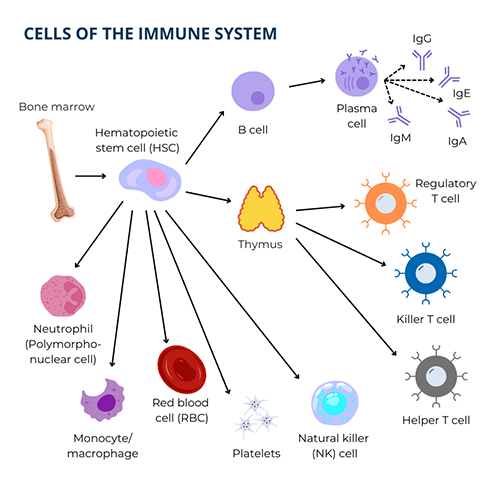
The main function of B cells is to make an important group of proteins called antibodies that recognize and physically attach, or bind, to germs. All together, B cells can produce a wide variety of antibodies against almost all germs. However, each individual B cell makes only one kind of antibody that recognizes only one specific type of germ, like a lock and key. For example, there are different antibodies, made by different B cells, that recognize chicken pox virus versus staph bacteria versus athlete’s foot fungus.
Importantly, B cells in the bloodstream become plasma cells, which are literally antibody factories that live mostly in the bone marrow but also in many tissues. One B cell makes one type of antibody and turns into a plasma cell that, in turn, makes large amounts of that same type of antibody.
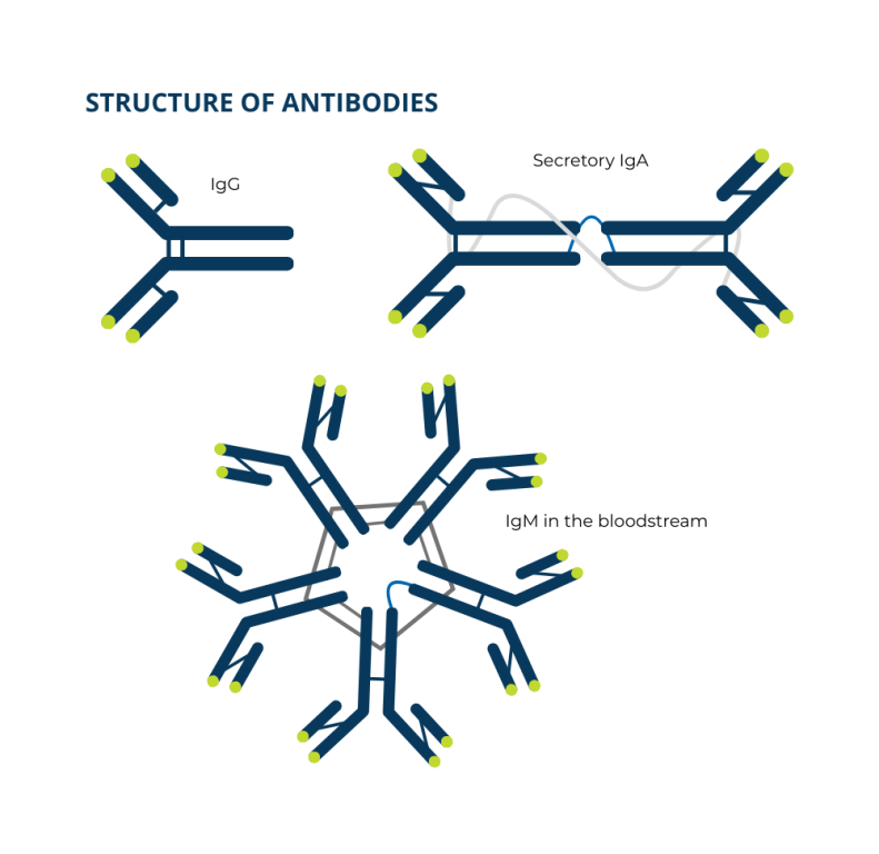
There are also different classes and subclasses of antibodies that differ from each other in their chemical structure. The five classes of antibodies are immunoglobulin (Ig) M, IgG, IgA, IgE, and IgD. IgG also has four different subclasses (IgG1, IgG2, IgG3, IgG4) and IgA has two subclasses (IgA1 and IgA2). Each class and subclass has a specific function in the body.
- IgM antibodies are the first class of antibodies B cells make and are important during the early days of an infection. IgM easily activates a group of immune system proteins called the complement system.
- IgG antibodies make up the majority of antibodies in the blood. They last for a few weeks and travel from the bloodstream into tissues easily. They’re also found in bodily fluids and only IgG antibodies cross the placenta from a mother to the baby. Immunoglobulin (Ig) replacement therapy contains mostly IgG. There are four IgG subclasses:
- IgG1: 60-70% of total IgG, protects against viruses and toxins from bacteria like diphtheria and tetanus.
- IgG2: 20-30% of total IgG, protects against the sugar coating (polysaccharide capsule) of certain bacteria like Streptococcus pneumoniae and Haemophilus influenzae.
- IgG3: 5-8% of total IgG, also protects against viruses and toxins from bacteria like diphtheria and tetanus.
- IgG4: 1-3% IgG4, controls immune responses.
- There are two subclasses of IgA antibodies, IgA1 and IgA2. IgA1 makes up about 80% of the IgA in the bloodstream. IgA1 and IgA2 in the bloodstream is called serum IgA and serum IgA is mostly single molecules. Both IgA1 and IgA2 in other bodily fluids like tears, bile, saliva, and mucus is called secretory IgA. There is a higher percentage of IgA2 in secretory IgA than in serum IgA. Secretory IgA is made up of two molecules joined together, which protects it from being broken down on mucous membranes and in bodily fluids. Secretory IgA protects mucous membranes in the respiratory and gastrointestinal tracts from germs.
- IgD makes up a very small percentage of antibodies in the bloodstream. Most IgD is on the surface of B cells along with IgM, and it may play a role in helping B cells develop into plasma cells.
- IgE antibodies protect against parasites and are also responsible for allergic reactions.
As part of their development in the bone marrow, B cells are tested to see if they make antibodies that could cause the immune system to attack healthy cells and tissues (autoantibodies). Any that do are weeded out. After developing, naïve B cells live in the bone marrow, lymph nodes, spleen, some areas of the intestines, and the bloodstream. These B cells make IgM and IgD and are called "naïve" because they haven’t yet come in contact with the germ their antibodies recognize.
- IgD and some IgM antibodies are anchored as single molecules on the surface of the B cells that make them.
- The other form of IgM is made up of five antibody molecules attached to each other in a ring and is found in the bloodstream.
After the IgM on a B cell comes in contact with the germ it recognizes, the B cell makes copies of itself. Some of the copies become long-lived memory B cells. Memory B cells form the ‘memory’ that allows for a fast, forceful response if the immune system sees the same germ again.
Other copies of the B cell ‘class switch’ to make other classes of antibodies, such as IgG or IgA, as they develop into plasma cells. However, regardless of the antibody class, the antibodies the plasma cells produce remain specific to the germ that was detected by the IgM on the original B cell.
Plasma cells are located in the spleen and lymph nodes throughout the body and pump out large amounts of antibodies, which end up in the bloodstream, tissues, mucus of the respiratory and gastrointestinal tracts, and other bodily fluids.
Sometimes, antibodies themselves prevent germs from causing an infection. For example, viruses must attach to and enter a person’s cells to multiply. Antibodies that bind to proteins on the surface of a virus can block it from entering cells and causing an infection.
In other cases, antibodies set off a chain of events involving other parts of the immune system that work to destroy the germ. Antibodies that recognize and attach to the surface of some types of bacteria activate the complement system, which then directly kills the bacteria. Antibody-coated bacteria are also much easier for white blood cells called neutrophils to eat and kill than uncoated bacteria. All of these actions prevent germs from successfully invading the body and causing infections.
How the immune system fights germs
Bacteria
Our bodies are full of bacteria, and bacteria are all around us on most surfaces. Our skin and the inner linings of our body, called mucous membranes, act as physical barriers that keep these bacteria from causing infections in tissues. But if the skin or mucous membranes are damaged by illness or injury, bacteria can get inside the body.
In most cases, bacteria are destroyed through the combined efforts of antibodies, complement proteins, and neutrophils. First, complement proteins and neutrophils act together to find and kill a bacterial cell. Then the B cell with IgM that recognizes that bacteria develops into plasma cells that pump out specific antibodies. The antibodies coat the remaining bacteria and activate complement proteins, which also coat the bacteria. The coating of antibodies and complement proteins helps neutrophils recognize the bacteria as foreign more easily.
The neutrophil starts its attack by attaching to the antibody and complement proteins coating the bacteria. It then stretches out around the germs and swallows them. Once the bacteria are in a pocket inside the neutrophil, special enzymes and toxic reactive oxygen species are released into the pocket, which kills the bacteria.
When antibodies, complement proteins, and neutrophils are working properly, this process usually kills the bacteria effectively. However, sometimes people get bacterial infections repeatedly, and these infections can harm tissues and organs. This can happen if there are too many bacteria or if there are problems with the production of antibodies, complement proteins, or neutrophils.
Fungi (yeast and molds)
Immune system defenses against fungal infections are complex and depend on the type of fungus. Candida albicans is a yeast-like fungus that commonly lives on our skin and mucous membranes without causing harm. A type of helper T cell called Th17 helps the immune system tolerate Candida in these areas while also keeping it from overgrowing and causing infections.
In contrast, molds like Aspergillus spp. are found mostly in the environment—especially in soil and air—and can cause infections when someone inhales their spores. The main defense against molds are neutrophils and macrophages, white blood cells that ingest and destroy fungal cells. However, T cells also contribute to antifungal defense by coordinating the immune response, especially if the infection is serious or invasive.
Viruses
We are often exposed to viruses. Our bodies defend against viruses differently than how they fight bacteria or fungi. Viruses can only live and grow inside our cells, which allows them to hide from parts of our immune system.
When a virus infects a cell, the cell sends out cytokines, especially interferons, to warn other cells about the infection. This warning usually stops other cells from getting infected. However, many viruses can get around this protection and continue to spread the infection.
T cells and NK cells that are moving around in the body are alerted to the viral infection. They travel to the location of the infection and kill the cells that are infected. This method of killing the virus is very destructive because many of our own cells are killed in the process. Even so, it is an effective way to get rid of the virus.
At the same time, T helper cells activate B cells, which begin making antibodies that recognize and block the virus. These antibodies help block infection if we're exposed to the same virus again. The immune system also makes memory T and B cells, which stay in the body and respond quickly if the virus returns—often preventing illness or making future infections milder.
Because neutrophils are not involved in fighting viruses, your white blood cell count usually is not high if you have a viral infection and may actually be low.
One of the biggest improvements in human health over the last 229 years has come from widespread use of vaccines [2]. Because of vaccines, serious viral diseases like polio, smallpox, measles, mumps, and rubella are not common anymore.
Understand PI more clearly

What is PI?
Also known as inborn errors of immunity (IEI), PIs are a group of more than 550 rare, chronic conditions where a part of your immune system is missing or does not function correctly.
Learn the signs and symptoms

Genetics and PI
PIs are mostly caused by changes, or variants, in genes that are important for how the immune system works.
Learn about genetics
Types of PI
There are more than 550 primary immunodeficiencies with distinct definitions, causes, and symptoms.
Explore PI types
This page contains general medical and/or legal information that cannot be applied safely to any individual case. Medical and/or legal knowledge and practice can change rapidly. Therefore, this page should not be used as a substitute for professional medical and/or legal advice. Additionally, links to other resources and websites are shared for informational purposes only and should not be considered an endorsement by the Immune Deficiency Foundation.
Adapted from the IDF Patient & Family Handbook for Primary Immunodeficiency Diseases, Sixth Edition.
Copyright ©2019 by Immune Deficiency Foundation, USA.
Sign up for updates
Receive news and helpful resources to your cell phone or inbox. You can change or cancel your subscription at any time.