

IDF logo
two older ladies sitting together and smiling

How PI affects other body systems

Key points

  • The immune system touches many different parts of the body. Oftentimes, the first symptoms of primary immunodeficiency (PI) show up as infections, autoimmunity, or inflammation in one or more body systems. 
  • Healthcare providers should test the immune system of anyone who develops unusual, severe, or multiple infections and/or inflammatory or autoimmune conditions to see if they have an undiagnosed PI.
  • Meningitis, encephalitis, and sepsis are conditions that people with many types of PI can develop that are medical emergencies. 
  • To check for eye problems, people with PI should get yearly eye exams by an eye specialist (ophthalmologist).
  • Sometimes, to treat inflammatory or autoimmune conditions in other organ systems, specialists may have to use immunosuppressants in a person with PI. Specialists should always work with the person’s immunologist to balance treating the condition with preventing infections.  

Because you need a working immune response to prevent and clear infections, people with primary immunodeficiency (PI) often have more frequent or severe infections than people without PI. In addition, people with many kinds of PI can develop conditions caused by an imbalance in the immune system known as immune dysregulation. One type of immune dysregulation is autoimmunity, where either T cells (autoreactive T cells) or antibodies from B cells (autoantibodies) target healthy tissue. Another type is chronic inflammation, where the immune system is generally overactive and healthy tissue becomes collateral damage.

Any organ or part of the body can be infected by germs, become the target of autoimmunity, or be damaged by chronic inflammation in someone with PI. Note that respiratory and digestive system symptoms are common across many types of PI, so they have their own pages. This page addresses the infections and autoimmune/inflammatory conditions most often seen in other body systems. 

Always tell your healthcare provider as soon as possible if you develop new symptoms that could be caused by an infection, autoimmunity, or chronic inflammation. All three can lead to serious, permanent organ damage if they are not treated. Some conditions, especially certain infections, can quickly become life threatening. It is important to see a healthcare provider that understands that people with PI sometimes do not have typical responses to infections. For example, those with PI may not spike a fever or have high white blood cell counts even if they have a severe and life-threatening infection [1].

Depending on your symptoms, your immunologist may need to work with other specialists to properly diagnose and treat your condition.

Nervous system

In people with many types of PI, serious infections like meningitis and encephalitis can develop within the nervous system. In addition, specific PIs, such as ataxia-telangiectasia (A-T) and many combined immunodeficiencies (CIDs), have neurological symptoms that are part of the overall condition. These can include difficulty walking straight (ataxia), cognitive impairments, attention-deficit hyperactivity disorder (ADHD), autism spectrum disorder (ASD), seizures, or inflammatory lesions in the brain, and are covered in the chapters specific to those PIs [2].

Meningitis 

Meningitis is inflammation of the lining of the brain and spinal cord (meninges). It is a serious and potentially life-threatening condition usually caused by bacterial, viral, or fungal infections. Note that drug-induced aseptic meningitis (DIAM), which sometimes happens as a side effect of intravenous immunoglobulin (IVIG) replacement therapy, is inflammation of the meninges that is not caused by an infection [3]. While DIAM is a serious IVIG side effect, it is not usually life-threatening and goes away on its own as few days after the infusion.

Symptoms of infectious meningitis include fever, severe headache, stiff neck, nausea or vomiting, and sensitivity to light (note that people with PI may not spike a fever in some cases). Meningitis is a medical emergency and people with these symptoms need to go to the emergency room right away. Emergency room providers will perform a physical exam and spinal tap (lumbar puncture) to test spinal fluid for the type of meningitis and the type of germ that may be causing it.

People with many different PIs are at risk of developing meningitis, but people with terminal complement deficiencies (C5-C9) or properdin deficiency are at particular risk for developing meningitis from Neisseria meningitidis bacteria [4]. This type of bacterial meningitis is especially dangerous.

Encephalitis

Encephalitis is inflammation of the brain itself. It can be caused by bacteria, fungi, or viruses (such as West Nile virus). It can also be caused by autoimmunity (autoimmune encephalitis). Symptoms include severe headache, confusion or altered mental state, fatigue, seizures, and a decrease in consciousness. Like meningitis, encephalitis is a medical emergency and people with these symptoms need to go to the emergency room right away.

To diagnose encephalitis, healthcare providers do blood tests such as a complete blood count (CBC), infectious studies such as spinal tap, magnetic resonance imaging (MRI), and neurologic examination. If an infection is causing the encephalitis, treatment includes treating the infection with antimicrobials. Treatment for both infectious and autoimmune encephalitis includes treating the inflammation with medications such as corticosteroids. 

Blood

Sepsis
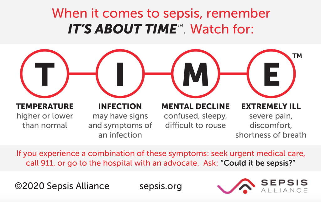
Infographic listing sepsis symptoms.
Developed by the Sepsis Alliance. 

Many different kinds of germs like bacteria, viruses, and fungi can cause a serious condition called sepsis, which is an extreme, whole-body response to an infection. Another, older term for sepsis is blood poisoning and it is still sometimes referred to as a blood infection. However, sepsis is not an infection, it is the body’s response to an infection. It is an extremely serious and life-threatening condition. Like meningitis and encephalitis, sepsis is a medical emergency that needs to be diagnosed and treated immediately.

Signs of sepsis include a high fever or chills, mental confusion or extreme sleepiness, disrupted vital signs like low blood pressure and high heart rate, and, in some cases, severe pain or discomfort (note that people with PI may not spike a fever in some cases) [5]. It can develop gradually or suddenly. Young children may refuse to eat and have low urine output. Septic shock refers to late-stage sepsis that includes very low blood pressure.

To treat sepsis, healthcare providers have to draw blood cultures to figure out what kind of germ is causing the underlying infection. Very often, the infection needs to be treated with intravenous (IV) antimicrobials even before an exact germ can be identified. 

Autoimmune cytopenias
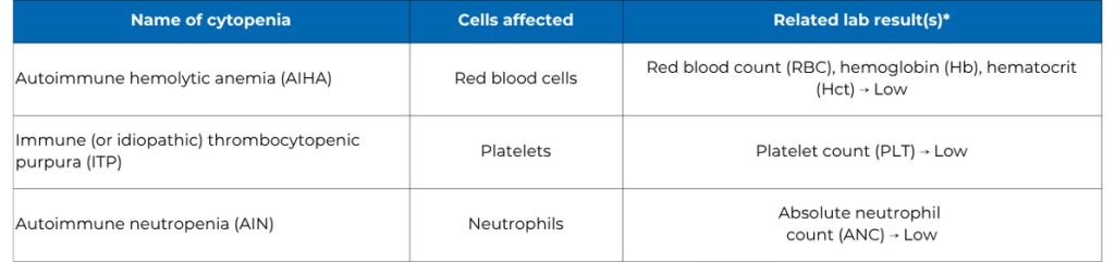
chart with cells affected by cytopenias
Autoimmune cytopenias, the cells they affect, and related laboratory test results. * These measures are part of the complete blood count (CBC) with differential blood test. Your healthcare provider may order additional tests to diagnose or track a particular type of cytopenia.

One of the most common types of autoimmunity seen across many types of PI is the development of autoantibodies that bind to and destroy healthy blood cells [6]. The blood cells affected can include red blood cells (RBCs), platelets, and white blood cells (WBCs). When autoantibodies destroy healthy blood cells, this leads to cytopenia, which is the general term for low numbers of blood cells.

Red blood cells (RBCs) use a protein called hemoglobin to carry oxygen through the bloodstream to all of the body's tissues. Anemia is when someone has a low number of RBCs or low hemoglobin so their tissues aren’t getting enough oxygen. There are different types of anemia. When the body makes autoantibodies that attack and damage the RBCs, this type of anemia is called autoimmune hemolytic anemia (AIHA). When the body has low hemoglobin, which is typically due to iron deficiency anemia, that is different from AIHA.

Symptoms of AIHA include feeling tired, having a headache, dizziness, fainting, and not being able to exercise as much. The person might look pale or ashen. If AIHA is severe, the person's skin and eyes can turn yellow, which is called jaundice. Their spleen may become larger than usual (splenomegaly) because it is trapping the damaged red blood cells. The body tries to make up for the lack of oxygen by making the lungs and heart work harder.

Trauma or damage to body tissues can cause bleeding. Platelets, which are small cell fragments, help form blood clots to stop the bleeding. A low number of platelets is called thrombocytopenia. When the body makes autoantibodies that attack platelets, causing thrombocytopenia, it is called immune (or idiopathic) thrombocytopenic purpura (ITP).
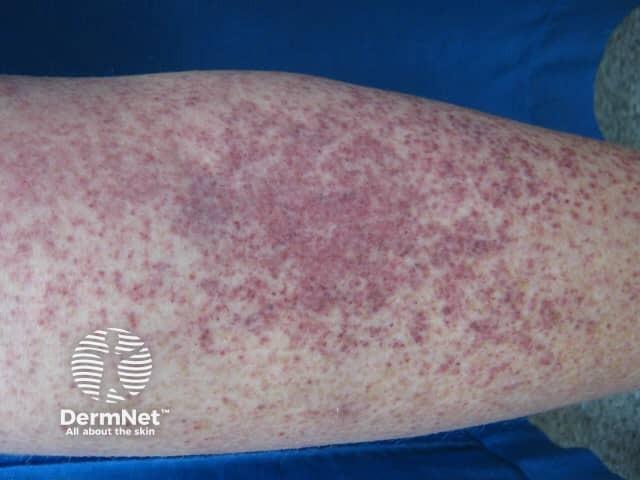
arm with skin rash
Example of petechiae due to thrombocytopenia. Image sourced from DermNet.

ITP can lead to severe or prolonged bleeding. People with ITP often notice more bruising than usual, sometimes in unusual places or without a known injury. They might have a pinpoint red rash called petechiae, which is caused by small bleeds under the skin. They may have nosebleeds that happen more often, last longer, and are hard to stop. Their gums may bleed easily. Their urine might be pink or red. Their stool may look black and sticky, like tar, which can mean there is bleeding in the digestive system. In rare cases, bleeding in the brain can cause confusion or even death.

Neutrophils are a type of white blood cell that help fight infections. When someone has a low number of neutrophils, it is called neutropenia. Autoimmune neutropenia (AIN) happens when the body makes autoantibodies that attack neutrophils.

Because neutrophils are first responders in the innate immune system, infections can get worse quickly in people with AIN. One of the most important signs of a serious infection in someone with AIN is fever (note that people with PI may not spike a fever in some cases). Other signs of infection, like coughing, throwing up, diarrhea, and rash, can also be present. Someone with AIN who has an infection may need to be checked in the emergency room or stay in the hospital. In these cases, they need antimicrobial medicines right away since the infections can be severe or lead to sepsis. People with AIN might also get ulcers or sores in their mouth and their gums may be red and swollen.

When a person has two or more autoimmune cytopenias, either at the same time or one after the other, they are diagnosed with Evans syndrome. Most often, those with Evans syndrome have a combination of ITP and AIHA [7].

Evans syndrome can occur in some types of PI like common variable immune deficiency (CVID) or autoimmune lymphoproliferative syndrome (ALPS), and there is evidence that it may often be the first sign of PI [8]. Evans syndrome can also occur when someone has a systemic autoimmune disease, such as systemic lupus erythematosus, even when these conditions are not related to a PI.

Diagnosing autoimmune cytopenias

Healthcare providers diagnose autoimmune cytopenias with blood tests. Usually, a complete blood count (CBC) with differential will show if someone has low numbers of red blood cells, different types of white blood cells, and/or platelets in their bloodstream.

To find out if the cytopenia is caused by autoimmunity, additional blood tests check for autoantibodies, although these antibodies are often difficult to detect. A specialist such as a clinical immunologist, hematologist, or oncologist typically evaluates individuals for these disorders. Sometimes, healthcare providers will do a bone marrow biopsy, which involves taking a sample of bone marrow tissue from the hip bone using a needle, to see if there is a problem with how the blood cells are made.

Treating autoimmune cytopenias

Autoimmune cytopenias may be temporary and if they get better on their own, they may not need much treatment. If they need to be treated, the goal is to get rid of the autoantibodies and allow the body to make more of the affected blood cell(s). The best treatment for a particular person depends on many factors and the particular type of cytopenia.

Treatment may include intravenous immunoglobulin (IVIG) therapy, corticosteroids, or other medications that suppress the immune system (immunosuppressants). Note that healthcare providers typically prescribe a higher dose of Ig to treat autoimmune cytopenias than to treat PI, so subcutaneous administration (i.e., SCIG) may not be an option.

In the past, healthcare providers regularly removed the spleen (splenectomy) in people with ITP. However, with newer therapies available, splenectomies are no longer the first line of therapy. In addition, since the spleen is an immune system organ, removing it increases the risk of severe infection in someone with PI, and should only be done with caution.

Autoimmune cytopenias often respond well to treatment. However, sometimes symptoms come back or need long-term treatment. People with autoimmune cytopenias rarely need blood transfusions unless their cytopenia is severe. In all cases, people with cytopenias need to be monitored regularly by their healthcare providers.

Most people with autoimmune cytopenias don't have to change their daily activities. However, people with low platelet counts may need to avoid activities with a higher chance of injury, like contact sports.

Endocrine system

The organs of the endocrine system release chemicals called hormones that are important for keeping the body working properly. The main endocrine organs are the pituitary gland in the brain, the thyroid and parathyroid glands, the pancreas, the adrenal glands, and the testicles (biological males) or ovaries (biological females). If the body makes autoantibodies that attack endocrine organs, it can cause major health issues. People with endocrine autoimmunity are often sent to a specialist called an endocrinologist to be diagnosed and treated.

Your thyroid is a small gland in the lower neck that makes hormones that are important for controlling how fast the body uses energy, along with growth and development. People that make too little thyroid hormone (hypothyroidism) usually gain weight, have a slow heartbeat, feel cold and tired, have trouble with bowel movements, and have rough hair and dry skin. On the other hand, people that make too much thyroid hormone (hyperthyroidism) usually lose weight, have a fast heartbeat, feel hot and anxious, and may have hair loss.

Sometimes, the body makes autoantibodies that attack the thyroid, causing inflammation (thyroiditis) that leads to either hypothyroidism or hyperthyroidism. Autoimmune thyroiditis, such as Hashimoto’s disease, is the most common autoimmune disease among the general population. In certain types of PI, including CVID and immune dysregulation, polyendocrinopathy, enteropathy, X-linked (IPEX) syndrome, autoimmune thyroiditis is even more common.

High blood sugar levels over a long period of time is a condition known as diabetes. Diabetes can happen when the body doesn't make enough of the hormone insulin (type 1) or when the body's cells don't respond to insulin (type 2). Autoantibodies cause type 1 diabetes by attacking the islet cells in the pancreas that make insulin. Once these special cells are destroyed, they don’t grow back. When a person loses too many of these insulin-making cells, they develop diabetes.

Type 1 diabetes is very common in some PIs, like IPEX syndrome, where it affects up to 80% of individuals [9].

The parathyroid gland controls calcium levels in the body. Autoimmunity against this gland is rare, but does happen in people with APECED syndrome. The parathyroid gland can also be underdeveloped in some PIs such as 22q11.2 deletion (DiGeorge) syndrome or CHARGE syndrome. The main symptoms of hypoparathyroidism stem from dangerously low calcium levels and can include muscle spasms, cramps, twitching, numbness or tingling of the hands or feet, and irregular heartbeat. If untreated, low calcium levels can lead to seizures.

Diagnosing endocrine conditions

Healthcare providers diagnose thyroid problems using blood tests that measure thyroid hormone. To see if a thyroid problem is caused by autoimmunity, providers look for certain autoantibodies in the blood.

Healthcare providers diagnose type 1 diabetes by testing a person’s urine and blood for a type of sugar called glucose. If blood glucose levels don't go down as they should after eating, or if they're high even when someone hasn't eaten, it could mean they have diabetes. Finding autoantibodies that target the islet cells can help prove that the problem is autoimmune.

Healthcare providers diagnose hypoparathyroidism by checking calcium, vitamin D, and parathyroid hormone (PTH) levels in the blood. Typically, an endocrinologist needs to evaluate someone for hypoparathyroidism.

Treating endocrine conditions

Most autoimmune endocrine conditions lead to a low level of essential hormones that are supposed to be produced by the endocrine organs. Treatment usually involves taking replacement hormones to try to reach normal levels. For hypothyroidism, extra thyroid hormone can be taken as a pill. Treatment for type 1 diabetes usually involves giving insulin through shots or an insulin pump. Although type 1 diabetes is caused by an autoimmune reaction, it is not yet clear whether using immunosuppressant drugs early in the disease changes the need for insulin treatment.

In the case of the thyroid, autoimmunity can also cause an increase in thyroid hormone, which requires removing or destroying at least part of the thyroid gland to fix the problem. This may require surgically removing part of the thyroid, radiation treatment of the thyroid gland, or using other medications. These treatments are always done under the care of an endocrinologist.

Treatment for hypoparathyroidism includes taking calcium and vitamin D supplements, along with dietary modifications. In addition, special precautions must be taken before surgical procedures to make sure the person’s calcium levels are normal.

Kidneys

The kidneys are part of the urinary system and have many tiny filters called glomeruli. The kidneys remove a waste chemical called urea from the blood. Urea is the main chemical in urine. The kidneys also control water levels in the body and levels of chemicals like sodium and potassium (electrolytes).

Glomerulonephritis is inflammation of the glomeruli, which can permanently damage them. When the glomeruli are damaged, the kidneys can’t filter the blood as well and urea, a waste chemical, builds up in the bloodstream. Kidney function gets worse over time if the inflammation is not treated. People with very low kidney function may need dialysis, which is regular filtering of the blood by a machine, or a kidney transplant.

Sometimes, especially in children, glomerulonephritis is caused by the immune system’s reaction to an infection like strep throat (post-infectious glomerulonephritis). However, it can also be caused by autoimmunity or chronic inflammation.

Autoimmune or inflammatory glomerulonephritis is the most common chronic kidney condition in people with PI. People with complement deficiencies, especially in components C1-C4, often develop autoimmune glomerulonephritis [10]. Autoimmune glomerulonephritis can also happen, but not as often, in other PIs like IPEX syndrome and STAT3 gain of function (GOF). People with Wiskott-Aldrich syndrome (WAS) can develop glomerulonephritis caused by a kidney disease called IgA nephropathy [11]. In IgA nephropathy, immunoglobulin A (IgA) builds up in the kidneys.

Diagnosing glomerulonephritis

Often, the first sign of glomerulonephritis is high blood pressure (hypertension). The person may have blood or protein in their urine, which can cause the urine to look brownish, like tea or cola, or foamy. If the person is losing a lot of protein in their urine, they may have swelling in their legs and feet from their body retaining extra water (edema).

Common blood and urine tests can help determine how well someone’s kidneys are working. The urine tests look for blood, protein, and white blood cells and measure electrolyte levels. If your kidneys are not working well, your healthcare provider will refer you to a specialist called a nephrologist for diagnosis and treatment.

The nephrologist may do a kidney biopsy to make the correct diagnosis. A kidney biopsy is usually done by inserting a needle through the skin and into the kidney to get a small piece of tissue. This tissue is then examined by a specialist called a pathologist, who performs tests on it, including looking at it under a microscope.

Treating glomerulonephritis

Typically, post-infectious glomerulonephritis does not require treatment with antibiotics since the infection may have already cleared. If the infection is still present, your healthcare provider may prescribe antibiotics. Your healthcare provider may also prescribe blood pressure medications to manage any hypertension.

To manage autoimmune glomerulonephritis, nephrologists often use medications that suppress the immune system (immunosuppressants). Because immunosuppressants weaken a person’s immune system further, they can make people with PI even more likely to get infections. To balance treating autoimmune glomerulonephritis with preventing infections, your immunologist and nephrologist need to coordinate these treatments together.

Joints and bones

As with other types of infections, people with PI can develop infections of the musculoskeletal system, such as septic arthritis and osteomyelitis. Septic arthritis is when there’s an infection of the joint space. It causes joint pain and swelling, and may also cause fever. Osteomyelitis is when there is an infection in a bone. Symptoms of osteomyelitis include bone pain, tenderness or a feeling of heat near the infected bone, as well as general infection symptoms like fever and chills.

To diagnose septic arthritis, healthcare providers use a needle to collect a sample of the joint fluid. They look for high numbers of white blood cells, as well as germs, in the sample. Sometimes, providers do blood tests such as CBC, blood cultures, erythrocyte sedimentation rate (ESR), and C-reactive protein (CRP) as well. Diagnosis of osteomyelitis includes similar blood tests but may also require imaging such as MRI. In some cases, a bone biopsy may be needed to make a diagnosis.

Both septic arthritis and osteomyelitis require antimicrobial treatment. Oftentimes, osteomyelitis requires several months of treatment. To help clear the infection, osteomyelitis may also require surgery in some cases.

People with particular types of PI develop septic arthritis or osteomyelitis more often than others. Antibody deficiencies, particularly X-linked agammaglobulinemia (XLA) and common variable immune deficiency (CVID), are linked to septic arthritis caused by several different types of bacteria [12]. In some cases, Mycoplasma spp. or Ureaplasma spp. bacteria cause the infection and can be difficult to diagnose because they are difficult to culture from patient samples.

Osteomyelitis can spread to the bone from the blood or develop from any deep infection that reaches the bone in a person with PI. However, those with chronic granulomatous disease (CGD) are at even higher risk of developing osteomyelitis [13]. In those with CGD, osteomyelitis is usually caused by bacteria like Serratia marcescens or fungi like Aspergillus spp.

Arthritis, or joint inflammation, is common both in people with PI and in people who don’t have PI. Different types of arthritis are named based on their root cause. Osteoarthritis is inflammation caused by joint ‘wear-and-tear.’ Rheumatoid, psoriatic, and juvenile idiopathic arthritis (RA, PA, and JIA, respectively) are conditions caused by autoantibodies attacking the joints.

People with PI don’t seem to have higher risk of osteoarthritis but some types of PI are linked to a higher chance of developing autoimmune arthritis. Unlike arthritis, myositis, which is muscle inflammation, is not very common in PI.

Many different antibody deficiencies have been connected to a greater risk of autoimmune arthritis [14]. One study found that about 15% of people with X-linked agammaglobulinemia (XLA) develop arthritis at some point [15]. People with CVID can develop rheumatoid arthritis or psoriatic arthritis, which also causes the skin condition psoriasis. These conditions can cause a lot of pain, limit daily activities, and potentially lead to lasting joint damage.

Common signs and symptoms of arthritis include joint pain and stiffness, swelling, and sometimes warmth or redness around the affected joints. Stiffness is often worse after not moving the joint, like in the morning or after resting, and it usually gets better with activity. When the arthritis is active, people may also have fevers, feel tired, and have a reduced appetite.

A physical exam by an experienced healthcare provider is extremely helpful in diagnosing arthritis. Your provider may refer you to a specialist called a rheumatologist for diagnosis and treatment.

Blood tests can help determine whether there is ongoing inflammation and look for specific autoantibodies, which may help determine what kind of arthritis someone has. X-rays, ultrasounds, CT scans, and MRIs of affected joints can show if there is active inflammation and if the joint has been damaged by arthritis. Sometimes, taking a sample of the fluid from inside the joint to test it can help determine the type of arthritis and make sure there is no infection in the joint. This is usually done by taking fluid out of the joint with a needle and syringe.

Treating arthritis often involves using medicines that weaken the immune system (immunosuppressants). Healthcare providers frequently first use steroids, like prednisone. These can be taken as pills, through an IV, or injected directly into the affected joints. They often work well for a while but can have serious side effects and do not prevent the arthritis itself from getting worse.

To better manage autoimmune arthritis, healthcare providers may use other immunosuppressants that aren't steroids. Because immunosuppressants weaken a person’s immune system further, they can make people with PI even more likely to get infections. To balance treating arthritis with preventing infections, your immunologist and rheumatologist need to coordinate these treatments together, and they may need to select therapies that don’t suppress your immune system as broadly.

Eyes and ears

Conjunctivitis, commonly known as pink eye, is when the eyelid lining and the white part of the eye (conjunctiva) are inflamed. Germs like bacteria or viruses, allergies, or things that irritate the eye like smoke or soap, can all cause conjunctivitis. Infection-related conjunctivitis can happen on its own or as part of other illnesses, like a cold. The usual signs of conjunctivitis are red and/or swollen eyes and eyelids, watery eyes, and mucus or pus leaking out of the eye. Pink eye can also make your eyes itchy, burn, or be sensitive to light.

In the morning, you might find your eyelids stuck together from dried mucus or pus that leaked from your eye while sleeping. Use a clean, warm, wet washcloth or cotton ball on each eye to loosen the dried material. After a few minutes, gently wipe each eye from the inner corner to the outer corner. It's important to wash your hands well afterwards, because pink eye infections are very contagious and spread easily.

You might need to see your healthcare provider if your vision is affected or if the pink eye doesn't go away after a few days so they can figure out what’s causing your symptoms. The provider will examine your eyes to see if one or both are affected and ask about your symptoms. If the provider thinks your symptoms are caused by bacteria, they may prescribe antibiotic ointment or eye drops. If it's not caused by an infection but by something irritating your eye, it's important to figure out what is causing the irritation so you can avoid it.

Some PIs can cause autoimmune eye conditions, where the immune system mistakenly attacks healthy eye tissue. Uveitis and iritis are two types of eye inflammation that can be caused by infection or autoimmunity. Uveitis is inflammation of the uvea, or layer of the eye between the retina and the white of the eye. Iritis, also known as anterior uveitis, is inflammation of the iris, or colored part of the eye surrounding the pupil.

Both uveitis and iritis may not cause any symptoms at first, so experts generally recommend that people with PI get yearly eye exams by an eye specialist (ophthalmologist) [16]. Note that it is important to see an ophthalmologist, not an optometrist, to check for these conditions. If inflammation in the eye goes untreated, it can lead to permanent vision loss. Treatment of autoimmune uveitis or iritis typically involves immunosuppressants such as systemic steroids or other medications.

Otitis media is a type of ear infection that happens in the middle ear, which is behind the eardrum and contains the three small bones (ossicles) that transmit sound to the inner ear. A small tube called the Eustachian tube connects the middle ear to the back of the nose and throat. In babies and young children, this tube is shorter and less slanted than in adults. This anatomy makes it easier for bacteria or viruses to enter the middle ear and harder for them to drain out. Sometimes, with infections or allergies, the Eustachian tube swells and closes, which stops the middle ear from draining at all.

The main symptom of otitis media is ear pain, often with a fever. This pain comes from irritated nerve endings due to inflammation or pressure changes. A baby or toddler may show they're in pain by crying, moving their head, or tugging at their ear. Older kids or adults might say the pain feels sharp. Other symptoms can include being restless or cranky, feeling nauseous, or throwing up.

Lying down can increase pressure behind the eardrum, which is why the pain is often worse at night. If the pressure gets too high, the eardrum can burst. If you see pus or blood draining from the ear, it could mean the eardrum has ruptured. Even though a ruptured eardrum can relieve pain, the infection is still there.

If you think you have an ear infection, it’s important to see a healthcare provider because repeated ear infections can cause scarring and permanent hearing loss. If the provider suspects a bacterial infection, they will prescribe antibiotics. They may also prescribe ear drops that help with the pain. You may need to go back for follow up to make sure the infection is gone and there is no fluid left behind the eardrum.

Healthcare providers may recommend a procedure called myringotomy with tympanostomy, or placing ear tubes, for children who get a lot of ear infections. An ear, nose, and throat (ENT) specialist (otolaryngologist) usually does this procedure. The ENT makes a tiny hole in the eardrum and puts a small tube through it. The tube helps fluid drain out of the middle ear and keeps the pressure even between the ear canal and the middle ear. The tube only stays in for a couple years and usually comes out on its own. 

Skin, teeth, hair, and nails

Some rare types of PI that affect parts of the body outside of the immune system, like Comel-Netherton syndrome, NEMO deficiency syndrome, or Job syndrome, lead to unusual teeth, fingernails, and/or hair. For example, people with these rare PIs may have dry, thin hair, teeth and fingernails that are not formed correctly, or a lack of sweat glands. These issues are part of the larger PI and are not caused by infections, autoimmunity, or inflammation.

In addition, one of the long-term side effects of hematopoietic stem cell transplant (HSCT) conditioning agents like busulfan is issues with the arrangement or size of teeth.

More commonly, people with PI have skin problems caused by infections, autoimmunity, or inflammation. Some of these skin conditions, like eczema or psoriasis, are common in people without PI too. A skin condition can be one of the first signs of a PI.

People with many types of PI can get skin infections caused by viruses, bacteria, fungi, or even parasites [17,18]. Common viruses that can cause skin infections include varicella-zoster virus (chickenpox and shingles), human papillomavirus (HPV; warts), herpes simplex virus (HSV; cold sores and genital herpes) and molluscum contagiosum virus. Bacteria like Staphylococcus aureus can cause pus-filled bumps under the skin (abscesses) and bacteria or fungi like Pseudomonas spp. can cause an infection of deep skin layers called cellulitis.

Particular skin infections are linked to specific types of PI in some cases. For example, chronic skin and mucous membrane infections from Candida spp. fungi (mucocutaneous candidiasis) are common in people with STAT1 GOF or APECED. Warts, hypogammaglobulinemia, infections, myelokathexis (WHIM) syndrome is part of a group of PIs that make people particularly vulnerable to warts and skin cancer from human papilloma virus (HPV). People with chronic granulomatous disease (CGD) can develop skin abscesses or cellulitis from several species of bacteria or fungi, including Serratia marcescens, S. aureus, Aspergillus spp. [19].

Eczema, also called atopic dermatitis, is a type of chronic inflammation of the skin. It is usually mild and is the most common skin problem in people with PI. It often starts as patches of dry, itchy skin that waxes and wanes in severity. People with PI who have other autoimmune or inflammatory conditions may also have eczema.

However, some types of PI cause more severe eczema. These include Wiskott-Aldrich syndrome (WAS), DOCK8 deficiency, IPEX syndrome, and some kinds of severe combined immunodeficiency (SCID). In these cases, the eczema may not respond to standard treatments such as topical steroid creams.

Psoriasis is an autoimmune skin problem that can be mistaken for eczema. The usual rash in psoriasis is called plaques. These plaques may be red, raised, itchy, and painful. These patches have a silvery scale on top. If the scale is removed, the plaque often bleeds. Psoriasis plaques are most common on the scalp, elbows, or knees. People with several types of PI, such as CVID, can develop psoriasis [20].

Several types of PI can cause the body to attack a person’s hair follicles, causing alopecia areata. People with alopecia areata develop bald spots because their body produces autoantibodies that target the cells, called hair follicles, responsible for hair growth. Alopecia areata specifically refers to circular areas of hair loss.

Additionally, some people develop vitiligo, a condition where the skin loses its coloring because the body produces autoantibodies that target the skin cells that make a pigment called melanin. The affected skin areas appear white, and how noticeable the change is depends on the contrast with the surrounding skin. The affected areas may change slightly over time.

Vitiligo and alopecia are most often linked to conditions like autoimmune polyendocrinopathy-candidiasis-ectodermal dystrophy (APECED), CVID, IPEX, and 22q11.2 deletion (DiGeorge) syndrome, but they can also occur in other types of PI.

Diagnosing skin and hair conditions

Often, an experienced healthcare provider can diagnose skin conditions with just a physical exam. If the skin looks unusual, the provider might need to do a skin biopsy. Healthcare providers usually take biopsies from the area where the symptoms are most obvious, using a razor to shave off the top layers of skin or a sharp tool to cut out a small, round piece of skin. This procedure is typically minor and can be done in the provider’s office with local numbing of the skin. Then, a pathologist will look at the skin sample under a microscope to figure out the type of cells that make up the rash. If the provider thinks you have a bacterial or fungal infection, they may order cultures from the skin sample to find out what kind of germ is causing the infection. 

Treating skin and hair conditions

Mild skin conditions can be diagnosed and treated by a primary care doctor or an immunologist. However, more severe skin conditions often require diagnosis and treatment by a skin specialist (dermatologist).

Many skin infections can be treated with antimicrobial medications specific to the germ causing the infection. Sometimes, people need intravenous antimicrobials if the infection is severe or deep. In the case of abscesses, a healthcare provider may need to drain the pus. Someone with severe cellulitis may need surgery to remove infected or dead tissue.

Autoimmune and inflammatory skin disorders are usually not life-threatening. However, they can cause significant emotional distress and, in rare cases, permanent changes in appearance. Severe rashes, like eczema, can allow bacteria and other germs to enter the bloodstream because the skin's protective barrier is damaged.

Treatment for most autoimmune and inflammatory skin conditions usually starts with applying moisturizing lotions and steroid ointments directly to the rash. If this is not enough to control the symptoms, stronger steroid creams or other immunosuppressant ointment medications can be applied. In rare cases, immunosuppressant medications may be needed to treat severe disease.

  1. Zhang S, Henderson TS, Scalchunes C, Sullivan KE, Jongco AM Iii. Persons living with primary immunodeficiency act as citizen scientists and launch prospective cohort body temperature study. J Particip Med. 2020;12: e22297.
  2. Xiao N, Huang X, Chen L, Zang W, Guan M, Li T, et al. Neurological complications in inborn errors of immunity: A scoping review of clinical spectrum, pathophysiological mechanisms, and therapeutic strategies. Clin Rev Allergy Immunol. 2025;68: 67.
  3. Lu Z, Lu X, Gao Y, Shang G, Zeng Y. Characteristics of adverse events and clinical risks of intravenous immunoglobulin: a pharmacovigilance study based on FDA Adverse Event Reporting System (FAERS). Front Med (Lausanne). 2025;12: 1724196.
  4. Ellison RT 3rd, Kohler PF, Curd JG, Judson FN, Reller LB. Prevalence of congenital or acquired complement deficiency in patients with sporadic meningococcal disease. N Engl J Med. 1983;308: 913–916.
  5. Symptoms. In: Sepsis Alliance [Internet]. 21 Jan 2019 [cited 29 Jan 2026]. Available: https://www.sepsis.org/sepsis-basics/symptoms/
  6. Schiavo E, Martini B, Attardi E, Consonni F, Ciullini Mannurita S, Coniglio ML, et al. Autoimmune cytopenias and dysregulated immunophenotype act as warning signs of inborn errors of immunity: Results from a prospective study. Front Immunol. 2021;12: 790455.
  7. Mathew P, Chen G, Wang W. Evans syndrome: results of a national survey. J Pediatr Hematol Oncol. 1997;19: 433–437.
  8. Hadjadj J, Aladjidi N, Fernandes H, Leverger G, Magérus-Chatinet A, Mazerolles F, et al. Pediatric Evans syndrome is associated with a high frequency of potentially damaging variants in immune genes. Blood. 2019;134: 9–21.
  9. Barzaghi F, Passerini L, Bacchetta R. Immune dysregulation, polyendocrinopathy, enteropathy, x-linked syndrome: a paradigm of immunodeficiency with autoimmunity. Front Immunol. 2012;3: 211.
  10. Zhang C, Liang D, Liu Z. Primary immunodeficiency as a cause of immune-mediated kidney diseases. Nephrol Dial Transplant. 2024;39: 1772–1784.
  11. Shimizu M, Kanegane H, Wada T, Motoyoshi Y, Morio T, Candotti F, et al. Aberrant glycosylation of IgA in Wiskott-Aldrich syndrome and X-linked thrombocytopenia. J Allergy Clin Immunol. 2013;131: 587–90.e1–3.
  12. Bloom KA, Chung D, Cunningham-Rundles C. Osteoarticular infectious complications in patients with primary immunodeficiencies. Curr Opin Rheumatol. 2008;20: 480–485.
  13. Winkelstein JA, Marino MC, Johnston RB Jr, Boyle J, Curnutte J, Gallin JI, et al. Chronic granulomatous disease. Report on a national registry of 368 patients. Medicine (Baltimore). 2000;79: 155–169.
  14. Pott NM, Atschekzei F, Pott CC, Ernst D, Witte T, Sogkas G. Primary antibody deficiency-associated arthritis shares features with spondyloarthritis and enteropathic arthritis. RMD Open. 2022;8: e002664.
  15. Hernandez-Trujillo V, Zhou C, Scalchunes C, Ochs HD, Sullivan KE, Cunningham-Rundles C, et al. A registry study of 240 patients with X-linked agammaglobulinemia living in the USA. J Clin Immunol. 2023;43: 1468–1477.
  16. Pham MN, Fuleihan RL, Sullivan KE, Cunningham-Rundles C. Ocular manifestations in primary immunodeficiency disorders: A report from the United States Immunodeficiency Network (USIDNET) Registry. J Allergy Clin Immunol Pract. 2022;10: 1788–1796.
  17. Zarezadeh Mehrabadi A, Aghamohamadi N, Abolhassani H, Aghamohammadi A, Rezaei N, Yazdani R. Comprehensive assessment of skin disorders in patients with common variable immunodeficiency (CVID). J Clin Immunol. 2022;42: 653–664.
  18. Al-Herz W, Nanda A. Skin manifestations in primary immunodeficient children. Pediatr Dermatol. 2011;28: 494–501.
  19. Leiding JW, Holland SM. Chronic granulomatous disease. GeneReviews(®). Seattle (WA): University of Washington, Seattle; 1993.
  20. Mormile I, Punziano A, Riolo CA, Granata F, Williams M, de Paulis A, et al. Common variable immunodeficiency and autoimmune diseases: A retrospective study of 95 adult patients in a single tertiary care center. Front Immunol. 2021;12: 652487.

This page contains general medical and/or legal information that cannot be applied safely to any individual case. Medical and/or legal knowledge and practice can change rapidly. Therefore, this page should not be used as a substitute for professional medical and/or legal advice. Additionally, links to other resources and websites are shared for informational purposes only and should not be considered an endorsement by the Immune Deficiency Foundation.

Adapted from the IDF Patient & Family Handbook for Primary Immunodeficiency Diseases, Sixth Edition 
Copyright ©2019 by Immune Deficiency Foundation, USA